Cerebrospinal fluid (CSF) drains from the skull through multiple pathways, some of which lead to cervical lymphatic vessels (CLVs) in the neck. We performed particle tracking velocimetry on recordings of fluorescent microspheres flowing through CLVs in living mice. Under control conditions (see first video), we found that fluid is driven primarily by the rhythmic contractions of the lymphatic vessels (so-called "intrinsic pumping"). However, following traumatic brain injury (see second video), the vessel contractions are disrupted and flow is greatly reduced. However, treatment with a norepinephrine antagonist after injury (see third video) large restores vessel contractions and fluid flow. Importantly, restoration of CLV drainage after traumatic brain injury led to reduced brain edema and improved cognitive outcomes for mice. From Hussain, Tithof, et al, Nature 2023.
The liver has arguably the most complex vasculature of any organ. It is also the only organ able to fully regenerate following resection. In collaboration with Drs. Tim Pruett and Joseph Sushil Rao, we have developed a lumped parameter network model which simulates blood flow through the entire human liver in just 12 minutes on a laptop. In the image to the left, we show how this model can be used to predict acute changes to hemodynamics following different surgical resections (colors indicate the volume flow rate of blood). From Tithof et al 2023.
Biofilms of Vibrio cholerae release adhesion molecules necessary for attaching to solid surfaces into their environment. This gives rise to a "public goods dilemma" related to the question of why exploitative "cheater" bacteria haven't evolved. With collaborators, we performed quantitative experimental analysis and numerical simulations of molecular transport in the vicinity of a biofilm. Our analysis resolves this dilemma by revealing that fluid flows characteristic of the Vibrio cholerae environment rapidly sweep away and dilute these adhesion molecules. From Tai, Mukherjee, et al 2022.
Perivascular spaces (PVSs) are annular channels surrounding the vasculature in the brain which provide a pathway for solute exchange between the cerebrospinal fluid (CSF) surrounding the brain and the interstitial fluid deep within the brain. We model the brain's PVSs as a branching network of hydraulic resistances, using published parameter values. A handful of parameters have wide uncertainties, so we run simulations for different parametric scenarios and test the simulation results against experimental observations. Our analysis suggests PVSs have low hydraulic resistance and the brain parenchyma (dense brain tissue) has high resistance. This scenario satisfies three empirical criteria: that the flow be driven by a small pressure drop, exhibit good CSF perfusion throughout the cortex, and increase substantially during sleep. Our results point to the most likely values of uncertain parameters, and they indicate which parameters should be measured in future experiments. From Tithof et al 2022; also see Boster et al 2022.
Recent studies show that impaired metabolic waste clearance from the brain plays a key role in the development of most cases of Alzheimer's disease (AD). It's also established that AD is correlated with reductions in sleep quality. An emerging picture suggests that with age, glymphatic clearance (which is most active during sleep) becomes impaired, playing a key role in AD etiology. We have simulated the competing effects of glymphatic clearance and aggregation of amyloid-beta (a metabolic waste molecule) into neurotoxic oligomers and plaques in a segment of brain tissue. These plots show predictions of the rate of plaque formation for Pe=0.9 (top) and Pe=3.6 (bottom). From Mukherjee & Tithof 2022.
Video file
Taylor dispersion refers to a process in which the combination of advection and diffusion leads to enhanced transport of a solute in a shear flow. Recent studies have suggested that a Taylor dispersion-like mechanism arising from an oscillatory flow (with zero net flow) may explain solute transport observed in perivascular spaces of the brain. We demonstrate that a steady flow component -- even in cases where it is much smaller than the oscillatory component -- leads to much greater solute transport. From Troyetsky et al 2021.
Remote video
oitadmin
Following a stroke, a wave of intense neuronal activity, called spreading depolarization, propagates through the brain (green wave). This wave causes arteries to constrict which pulls an abnormally large amount of cerebrospinal fluid into the brain, causing swelling. From Mestre et al, Science 2020.
Video file
A model of perivascular spaces in the brain which we used to demonstrate that a moderately elongated shape permits greater fluid flow than a circular annulus. The “optimal” shape that we compute is very close to that of real perivascular spaces observed at the surface of the brain in mice and humans, which is perhaps a consequence of evolutionary optimization. Read more in Tithof et al, Fluids Barriers CNS 16 (2019): 19.
Video file
By injecting 1 µm spheres (green dots) and performing particle tracking velocimetry, we obtain quantitative measurements of cerebrospinal fluid flow through perivascular spaces in living mouse brains. Flow speeds are slower for high blood pressure (right panel) than normal blood pressure (left panel). Based on results reported in Mestre, Tithof, et al, Nature Commun. 9 (2018): 1-9.
Video file
The glymphatic system may offer a promising novel route for drug delivery to the brain. Here we inject a living mouse with hypertonic mannitol and image cerebrospinal fluid influx throughout the entire brain (left). Front tracking velocimetry (right) allows us to quantify the influx area and speed. From Plog et al. JCI Insight 3.20 (2018): e120922.
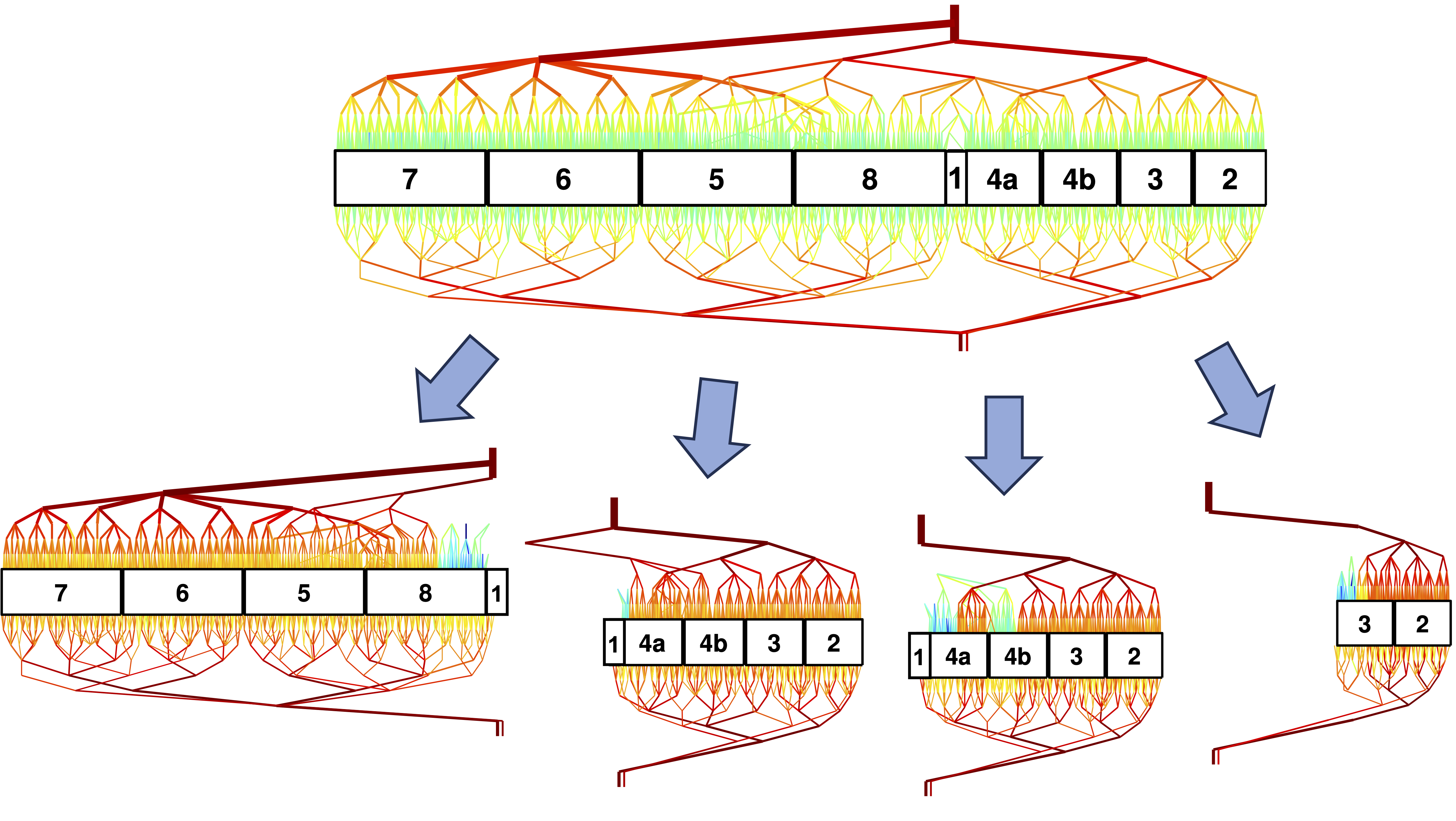
A state space projection showing the evolution of a chaotic Kolmogorov-like flow (blue curve) as it visits the neighborhoods of unstable equilibrium solutions (gray and red spheres). From Suri, Tithof, et al., Phys. Rev. Lett. 118, 114501 (2017).